


Written by Sajid, Health Content Researcher
A visual look at what cankles can look like, with brief context on common causes. For informational purposes only, not a substitute for professional medical advice.
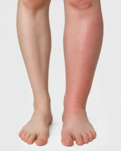
These cankle pictures show what fluid related swelling in the lower leg can actually look like, shown next to a normal, unaffected leg for comparison. Not every cankle looks like this, plenty of people simply have a natural leg shape with no swelling at all, but the pictures below focus on the visible, swelling related kind people most often search for.
The first image above the title shows a clear side by side difference between a normal calf and one affected by swelling. The right leg shows redness and a rounded, puffy shape that covers the ankle bone completely. This kind of visible swelling is usually fluid related rather than fat or bone structure. Warmth and redness like this can point to inflammation or venous insufficiency, a common circulation related cause. Sudden swelling that looks like this is worth mentioning to a doctor rather than waiting it out.
This image shows a closer view of one swollen leg next to a normal one, with the ankle bone barely visible on the affected side. The skin looks stretched and slightly shiny, a common sign of fluid pressing against the surface from underneath. This kind of swelling tends to build up gradually rather than appear overnight. Standing for long periods or a condition like lymphedema can both create this look. Comparing both legs side by side like this is often the easiest way to actually notice the difference.
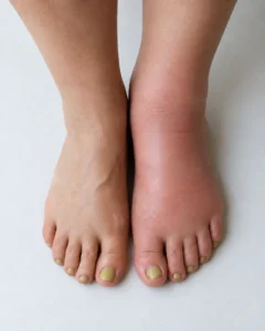
This picture shows a top down view of both feet, with one side clearly more swollen than the other. The loss of visible tendons and bone structure on the swollen side is a common sign of fluid retention. One sided swelling like this generally deserves more attention than swelling in both legs, since it can point to a localized issue. This is one of the clearer cankle pictures for seeing how much shape can actually change with swelling. A doctor can help confirm whether an image like this reflects something that needs treatment.

This image shows swelling concentrated mainly around the ankle and lower calf, while the foot itself looks closer to normal. This kind of pattern is common with conditions that affect circulation rather than the whole limb evenly. The visible color change also shows how blood flow and fluid pressure can affect skin tone. Swelling that stays in one specific area like this is sometimes easier to treat once the cause is identified. Pictures like this help show why the location of swelling matters, not just that it is present.
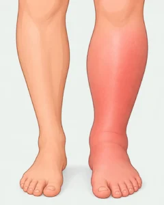
This illustration gives a simplified look at what is actually happening beneath the skin during ankle swelling. The shaded area represents where fluid has built up in the tissue, softening the natural curve of the ankle. Illustrations like this help explain the mechanism, since real swelling can look a little different from person to person. Illustrations like this pair well with real cankle pictures to explain why the leg changes shape the way it does. Think of it as a reference point, not a diagnosis.

This picture shows a more real world example of leg swelling, with both feet elevated during a checkup. Elevating the legs like this is a common first step used to see how swelling responds to position. This angle differs from the standing photos above, since it shows how the legs can look while resting. Comparing swelling in different positions is part of how doctors and patients track whether it is improving. Pictures like this show what an actual evaluation for cankles can look like.
Not every cankle looks like the swelling shown in these cankle pictures. Many people have cankles that are simply their natural shape, with no redness, warmth, or puffiness involved at all. If you are trying to figure out whether your own legs are swelling related or just structural, that comparison walks through the difference in more detail.